From Stem Cells to Organs - The Growth of a Human Being
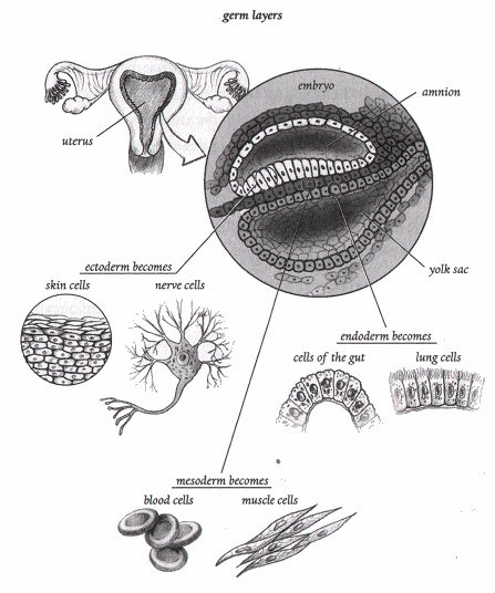
At about two weeks, the embryo enters a new phase that signals the start of a body blueprint1. Cells from
the ICM (inner cell mass) change and organize into three kinds of cells called germ layers2, illustrated
above. Cells from each germ layer begin to further develop into tissues and organs of the body (only major
cell types and structures are listed):
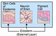
o Cells of the ectodermal3 layer become nerve and skin cells and form the inner ear, eye, mammary glands
(Milchdrüsen), nails, teeth, and the nervous system, including the spinal cord (Rückenmark) and brain.
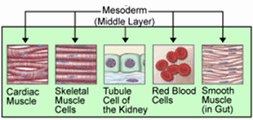
o Cells of the mesodermal3 layer become blood, muscle, and bone cells, and make the heart, skeleton,
gonads Keimdrüsen), urinary system, fat, and spleen (Milz).
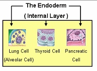
o Cells of the endodermal3 layer become cells inside the gut (Darm), liver, pancreas (Bauchspeicheldrüse),
bladder, lungs, tonsils (Mandeln), pharynx (Rachen, Schlund), and parathyroid glands (Nebenschilddrüsen).
At one month of age, the human embryo is three millimeters long, roughly the size of a pea. Now cells rarely
function as individual actors; they start to work in concert with their neighbors. During this time, a
primordial4 heart begins to beat. In a few days, it will pump blood through a graceful
triad5 of circulatory arcs. Even the embryonic heartbeat, without a functioning
system of blood vessels, is essential to development. Recent research in zebrafish completed at the
University of California, San Francisco, has found that the very action of a beating heart is a signal
for the formation of proper functioning valves. Apparently, blood flow sets certain cells on the
trajectory to become part of a heart valve6. If true, this information could lead to the
prevention of in utero defects7 caused by the interruption of the embryonic heartbeat in humans.
The early days are crucial. Signals between cells increase, causing more cells to differentiate. The
basic form of the body is laid down when, along with a primordial heart, the embryo develops structures
that will become the future spinal cord and brain and small protuberances8 (called
limb buds) where the limbs will be. The optic vesicle9, a slight depression where the eye
and optic nerves will grow, rests in a cusp along the mushroom-like swelling that will become the
cerebrum10. The embryonic stem cells have done much work in just a few weeks.
During the next four weeks, all the major organ systems appear. The embryo changes from a beanlike
crescent11 to a form with undeniably human characteristics. At the end of two months, the
embryo has recognizable limbs and a heart, along with a brain, eyes, ears, and a nose. Perhaps the
most astonishing accomplishment during this phase is the formation of the nervous system. Neural
anatomy is not a single organ - it is an intricate system of organs and conduits12
routed throughout the body. Following the fate of embryonic stem cells along one pathway in the
laboratory shows how convoluted13 their journey can be.
The outermost germ layer, the ectoderm, eventually becomes part of the body's entire sensory and motor
apparatus. An opening cascade of cell signals causes the topmost portion of the ectoderm to further
diversify into specialized cells of the nervous system. Brain-related structures must be planned and
executed simultaneously by other embryonic cells. For example, the brain needs a container to grow in,
so genes in a different layer, the mesoderm, must coordinate the development of the skull. Other genes
must coordinate the pace of growth so that the skull will develop at the same rate as the brain. As
they gain mass through cell division, the brain and skull will require the support of muscles and
tendons14 formed by different kinds of mesodermal cells.
c. 620 words
Source: Stem Cell Now by Ch. Th. Scott, A Plume Book, USA, 2006, pp. 29-31
|